Published on
How zebrafish gained their popularity as a model organism
By: Samantha Kummerer, Bond LSC
The core of many modern discoveries in developmental biology is swimming in a tank.
These are zebrafish that serve as the lab rats for Anand Chandrasekhar’s research.
Dozens of tanks containing thousands of swimming fish fill the lab in the basement of the Bond Life Sciences Center. There are baby fish, striped fish and clear fish, many genetically modified for experimental reasons, and they are studied from fertilized embryo to adolescence.
Chandrasekhar’s lab uses zebrafish to study migrating motor neurons in the brainstem that control muscle movement in the face and jaw.
Multiple types of neurons positioned precisely throughout the brain connect together to form networks. Those networks give the brain its ability to function.
“For us, it is important to study how these networks form because the brain is what makes us human,” said the Bond Life Sciences Center researcher.
During development, neurons respond to signals that enable them to move to different locations and form networks, Chandrasekhar explained. If the neurons don’t migrate properly to specific locations then the brain can’t function properly.
It is a domino effect. If the brain stem motor neurons don’t migrate correctly then the corresponding neural networks don’t form correctly and then the fish are not able to eat well.
Chandrasekhar’s lab is captivated by this migration. Different neurons move different ways and are set into motion by different signals. What are the signals to tell the neurons to stop or to go? Do these signals vary based on neuron type and species?
The lab also seeks to understand the repercussions of deficient migration using genetically modified fish.
Cell migration doesn’t just occur in the brain or with nerve cells. Cells also need to move in particular ways to form the heart and to fight infections.
A Model Swimmer
The zebrafish has long joined traditional lab rats and mice as scientists’ choice as an ideal test subject.
“When I first started out I wasn’t entirely sure I could do that (work with zebrafish), because how am I expected to handle something in water, moving around and be able to use it to really do experiments?” Chandrasekhar said. “But once you see how you handle fish, how you get them together, it just becomes one more thing that you do and then it makes you wonder how people work with mice.”
Like other model organisms, zebrafish share many genetic similarities with humans. These similarities mean researchers can investigate cancer, heart disease, muscle and tissue disorders in humans by testing and studying fish.
Mice and rats also share a large portion of DNA with humans, but the zebrafish’s unique characteristics enable experiments and observations to be cheap, efficient and fast.
One of those unique traits is a rapid development rate. This allows researchers to study important developmental stages in a single week.
“They’re incredibly efficient in terms of, I can set them up and they’ll be ready tomorrow morning,” said Ph.D. student Devynn Hummel.
The fact that embryo development in mice occurs inside their mother also makes observing early stages of development hard. Zebrafish embryos, on the other hand, grow independently, outside the mother.
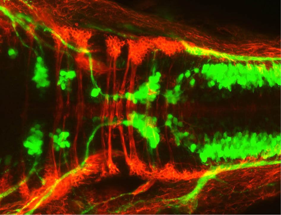
Neurons in the hindbrain of a zebrafish embryo are illuminated due to the use of florescent chemicals. The branchiomotor neurons are labeled with green and the commissural axons are labeled with red. | Photo by Suman Gurung
The young zebrafish’s transparency also helps researchers track cells within its body.
Hummel explained that by using fluorescently tagged molecules we are able to zero in on anything from neurons to changes in calcium concentrations, allowing fish to be used in a wide range of different studies.
“We’ve sort of engineered them to allow us to look at different tissues,” Hummel explained. “It really is remarkable the details in imaging zebrafish and what you can see. It’s truly extraordinary.”
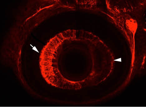
An eye of a zebrafish is illuminated 48-hours after fertilization using immunohistochemistry.| Photo by Suman Gurung
Scientists throughout the world are catching on to the advantages of the fish.
Chandrasekhar said there are significantly more zebrafish labs throughout the world than there were 20 years ago.
Despite its advantages, the zebrafish will not completely replace mice as a lab tool.
Chandrasekhar explained there are still certain experiments where mice are more advantageous. The same area of research can be explored using zebrafish, mice or even fruit flies, but the specific questions you ask change based on the tools available.
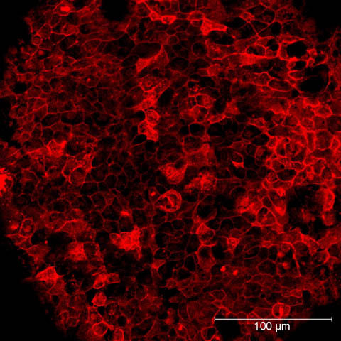
A snail toxin, MVIIA, is illuminated in a zebrafish embryo. The toxin blocks calcium channels needed for neurotransmitter release. Chandrasekhar’s lab studies motor neurons using zebrafish. | Photo by Devynn Hummel
Beyond Nerves
While Chandrasekhar’s lab concentrates on neurons, understanding how the migration occurs can be applied in a different context because of shared cellular mechanisms.
“Some of these molecules are just tools that different types of cells can use in different ways to accomplish different functions,” he said. “You can study a steering wheel in a car and know that it’s allowing the wheels to turn and the same steering wheel in a truck is allowing the wheels to turn in a truck, but maybe at a different time and a different way.”
Members of Chandrasekhar’ lab already experienced a broader implication of their research when a gene they studied with a role in neuron migration provided insight on Spina bifida, a failure of the fetal spinal cord to close completely.
“We all hope the problem we study has a broader impact on human biology so there’s always a quest to dig deeper and learn something new,” Chandrasekhar said.
The lab is hoping one day soon this fishy tool will help shed even more light on the mechanisms maintaining human health. For now, the researchers and their fish will just keep swimming.
With more than 2,000 fish, the lab has no plans on slowing down.
Anand Chandrasekhar is a Biological Sciences professor at the University of Missouri. He uses mice and zebrafish to study the mechanisms involved with the development of the nervous system. His lab in Bond LSC uses cell biological and genetic methods to understand these mechanisms. He received his Ph.D. in biology from the University of Iowa.