Published on
It’s an asset to be able to visualize and think about the nervous system from the perspective of an electrical engineer.
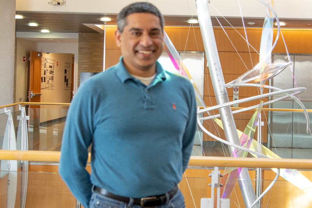
Cell biologist Anand Chandrasekhar — whose work focuses on the movement of neurons within the brainstem of mice and zebrafish, as well as on the consequences of that movement or lack of movement for the animal’s behavior— brings that angle to his work all the way from his undergraduate degree in electrical engineering he received in his native country of India. Although much of the nitty gritty details of his engineering knowledge have been lost to time, the legacy of those concepts has stayed with him in his neurological research at MU’s Bond Life Sciences Center.
“I’ve always been interested in seeing how things work and how processes are connected, and I think that’s because of my engineering background,” he said. “The nervous system was a natural thing to study for me because I’ve always been curious as to how circuits work within an organism’s brain and how those circuits allow or disallow certain biological processes to happen.”
That circuit-related curiosity led to Chandrasekhar’s two decades-long investigation of the movement of neurons within the brain.
“Neurons are not just in a spot in the brain from the beginning,” he said. “They are told to move there at some point in the embryonic stage. If they do not move to the proper spot within the brain, the consequences can be quite severe.”
He uses mice and zebrafish for his experiments because they are both vertebrates, meaning they have a lot in common with the human brain in structure and function. Previous scientific research shows when neurons don’t move properly within a human brain the consequences are devastating, resulting in a spectrum of cognitive and motor disabilities. In severe cases, the individual does not survive to birth. So, the consequences to the animal of neurons that don’t move to their rightful place during development can be quite dire.
Two projects related to neuron movement take up the bulk of the Chandrasekhar lab’s time, with graduate students Emilia Asante and Devynn Hummel diligently studying each. Asante’s project focuses on zebrafish, and looks specifically at the consequences for the fish when neurons in the brainstem do not migrate properly. “We have some understanding what the actual consequences of defective neurons are in humans, but it’s not so clear with fish,” she said.
Zebrafish embryos and larvae are transparent, and Asante takes advantage of this fact with her work. For one particular series of tests, Asante feeds the larval fish tiny fluorescent particles so that she can tell how much food the fish eat when she puts the live fish under a microscope. Compared to wild type (normal) fish, the mutant fish (those with defective neuron movement) ate less food than their normal counterparts.
Armed with this conclusion, Asante took her work a step further, and she asked why the mutant fish didn’t eat as much. So, she designed a test to look at the jaw movement of the zebrafish to investigate whether that had something to do with their reduced food intake. Asante devised a method such that fish could move their head but were not able to swim away. This allowed her to record the jaw movement of the fish. Asante found that the fish who did not eat as much as others were, by and large, the same fish whose jaw movement was less frequent. At the same time, Asante observed that the fish whose jaw movement was reduced were normal in every other way, including their ability to swim and capture food. Thus, Asante identified slowed jaw movement as the definite effect of defective neuron movement.
Now, Asante is investigating what upstream or downstream issues could cause this reduced jaw movement.
“In science, processes often don’t work in isolation,” she said. “So, there could be any number of things that are causing the fish to have slower jaw movement. They may not be getting a signal from their brain that tells them they’re hungry. Even if they do get that signal, they may not be able to move it downstream and tell their jaw to move so that they can chew on, swallow and eventually digest as much food as they need.”
Hummel is taking his investigations in a different direction. He works with mice and is currently studying how a set of genes functioning within the brainstem interact with each other in order to allow for proper neuron migration. Hummel’s work is focused on how cells move in general, in contrast to Asante’s. However, Hummel’s work still uses the neurons that control the jaw as a model cell type. As is the case with zebrafish, neurons that don’t migrate properly can cause severe behavioral defects in mice.
Several years ago, the lab discovered a gene that controls the direction in which neurons migrate, as compared to other genes required for the ability to migrate.
“This is exciting, because such a gene had not been previously identified,” he said.
Unlike the zebrafish that Asante works with, Hummel must dissect pregnant female mice in order to determine the extent of neuron migration within a developing embryo. This means tracking pregnant females over several days prior to ever beginning an experiment. Managing a colony of over 200 mice is no easy task, yet Hummel stays motivated since his work can yield such meaningful results.
“One of the coolest things about doing this kind of work is that sometimes, no one in the world has ever investigated specifically what you are looking at,” he said. “By that criteria, once you make a discovery, you know more about that subject than anyone else in the world.”
Chandrasekhar agrees that seeking answers to these big questions is what makes his work so fulfilling.
“Neuron movement within the brain is similar across the board – in humans, mice and zebrafish,” he said. “If we can understand how these cells move — as our work with mice tries to figure out — or why they move the way they do — as our work with zebrafish tries to figure out — in simple systems, the hope is that we can then transfer that knowledge to improve our understanding of the human brain.”


