Published on

By Lauren Hines | Bond LSC
At 24 weeks pregnant, a baby can hear the mother’s lullabies. At 30 weeks, her belly is a little over a foot large. At 40, the hospital bag is already packed and ready to go.
But imagine delivering only two weeks after the bump starts showing.
Preeclampsia makes induced birth necessary as life-threatening symptoms start 20 weeks into pregnancy, and delivery is the only cure. There is a lack of ways to detect it, and it’s difficult to ethically study the early stages of human reproduction. But what if it was possible to rewind the process to see when the source of the disorder took hold?
Different cellular models of the placenta might be that time machine researchers need to study early pregnancy disorders.
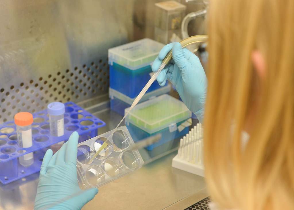
The Michael Roberts lab at Bond Life Sciences Center combines the knowledge and practices of three cellular models to learn more about early pregnancy and diseases.
“We can use these different models to study placental infection with viruses like Zika virus and, of course, COVID because there’s still a controversy as to whether COVID is a hazard in early pregnancy,” principal investigator Michael Roberts said. “And my suspicion is it probably is.”
The placenta is the brand-new organ generated by the embryo — the baby — before any of its other organs develop to give the baby nutrients and support as the embryo grows.
Megan Sheridan, postdoctoral fellow working with the Roberts lab, works on a project to combine 2D and 3D models to see how Zika and Dengue virus interact with the placenta and affect early pregnancy.
“[Complications] occur but you don’t necessarily know that until the end of pregnancy when the baby is delivered,” Sheridan said. “We really want to kind of go back in time and try to determine what’s going wrong in early pregnancy.”
Modeling early placental development is vital to see the beginning of disease complications, but the challenge is to get an accurate glimpse while not putting a healthy pregnancy at risk.
“It’s hard to get a good model to do research on that,” said Jie Zhou, postdoctoral fellow in the Roberts lab. “So that’s why we are working on different stem cells and trying to build up the best model to work on.”
So, the Roberts lab uses the BAP model. By working with a hospital, the lab first takes fibroblast cells from discarded umbilical cords after the baby is born. These fibroblast cells can be collected from mothers experiencing a normal pregnancy or pregnancies associated with complications, like preeclampsia. Then, as Sheridan puts it, they add a “cocktail of genes” to turn the cells into induced pluripotent stem cells. From there, the lab adds BAP — a mixture of growth factors and inhibitors that turn the stem cells into trophoblast cells.
Now the lab is back at the beginning of pregnancy except in a Petri dish of cells. In pregnancy, these trophoblast cells line the outside of the embryo.
From here, researchers can see what possibly caused the pregnancy complications in the first place.
“It’s trying to mimic pregnancy in a dish, essentially by using cells that will develop into placental-type cells,” Roberts said.
However, a flat Petri dish is nothing like a real placenta.
“It’s two-dimensional, and we can only culture the cells up to eight days after treatment so if we want to do long-term experiments, we can’t use this model,” Zhou said.
Two alternative models came out in 2018. The first model was by Hiroaki Okae et. al who created a 2D trophoblast cell line where trophoblasts can continually reproduce themselves long past eight days. The second model by Margherita Turco et. al also created a similar cell line, but in three dimensions.
This last model inches even closer to an early placenta. These 3D organoids float around inside a jello-like substance called Matrigel where they self-organize into a placental-like structure.
Together the two models give them a full picture. The 3D model gives a clue into how multiple cell types interact with each other while the 2D model allows researchers to see how a single cell type responds.
Since no model is perfect, the Roberts lab is combining their BAP model with protocols and growth conditions from the other two models to create a foundation for their experiments. Now researchers can start asking deeper questions.
While nothing is quite like a real womb, the Roberts lab will continue working backward.
“All of these models, put together, are really useful because we can kind of use them to their fullest potential and systematically assess which one might represent a certain disease or best answer a certain research question,” Sheridan said.