Published on
Updated on
How advanced light microscopy brings clarity to research questions
By Sophie Rentschler | Division of Research
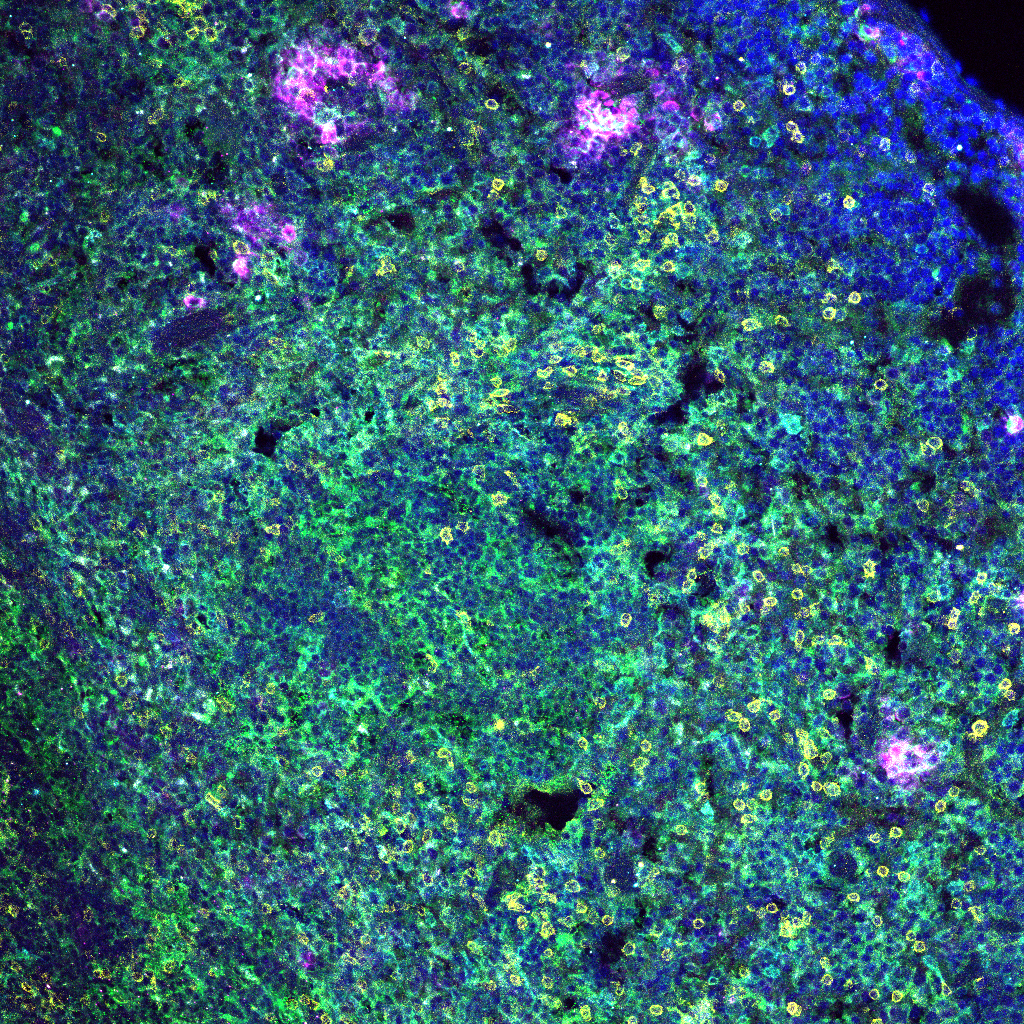
Under the microscope, the slice of mouse organ tissue resembled a mix of swirling indigo sky from Van Gogh’s Starry Night mixed with one of Monet’s impressionistic water lily paintings.
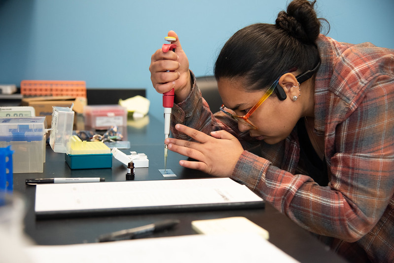
While it could be mistaken for abstract art, this high-definition picture of cellular interactions and distinct structures tagged with bright, highlighter-esque colors are the exact images Paul de Figueiredo needs to explore a form of cancer treatment.
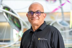
“Our eyes are one of our major senses,” said de Figueiredo, a Bond Life Sciences Center principal investigator and professor of Molecular Microbiology and Immunology. “Imaging is our way of learning about biology.”
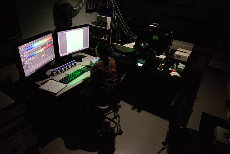
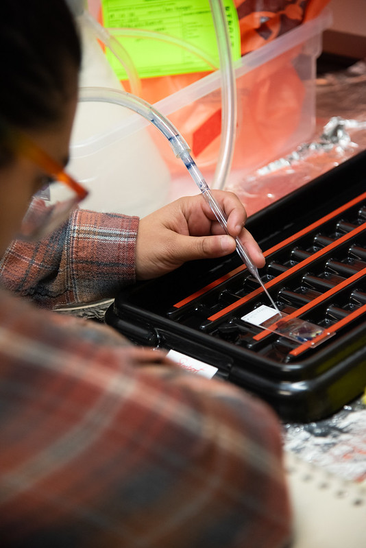
His work relies on collecting this cellular information in the right way at the right time, and that examination begins with experts like Tara Finegan, director of the Advanced Light Microscopy Core. Instead of conducting its microscopy in-house, the de Figueiredo lab utilizes the ALMC’s full-service imaging to take samples from preparation to final picture using equipment like the core’s Leica SP8 confocal microscope.
The cost for the ALMC’s high-quality images is considered cheap within the light microscopy market, so the de Figueiredo lab uses its confocal and wide field microscopes most.
“A Formula 1 car is expensive because of the highly sophisticated technology; ALMC’s images are really good but still cost-effective and reliable,” he said. “The cost of imaging would be five times more through a private company.”
In the last fiscal year, 238 people utilized the core’s services as part of their research initiatives. It saw a 28% increase in usage compared to the previous year.
Finegan sat in her office where a flyer on the wall represents the compromises that come with microscopy. The flyer follows her to consultations with researchers and department retreats, demonstrating what kinds of imaging you can get with each service at the core.
“If you want higher resolution images, then you compromise the speed of the imaging process,” she said.
While the ALMC’s confocal microscopes are the most used piece of technology — these workhorses making up 56% of all services in fiscal year 2025 — a free consultation with her staff helps home in on the optimal service the core can provide for each lab and the type of research at play.
A microscope for every occasion
The core has a handful of different scopes: stereoscopes, widefield, spinning disk confocal, point scanning confocal and super-resolution microscopes. Each instrument has a specialty.
Stereoscopes are very fast but lack sensitivity. These scopes are frequently used to screen genetically modified plants, gathering a quick and 3D image of organisms.
The widefield’s most common use among ALMC clientele is taking pictures of slides. Its resolution can be blurry if samples are not super-thin, but can be helpful to researchers to make quick conclusions about their samples.
To watch a live, intercellular event, Finegan recommends the spinning disk confocal. Researchers can watch sub-cellular interactions at high resolution, and even process data to acquire super-resolution images.
The confocal scopes can image molecular complexes and sub-cellular interactions through large tissue structures. Super-resolution capabilities on these scopes can allow for nanoscale resolution. Advanced techniques can take hours to set up, let alone produce images. But, the super-resolution stimulated emission depletion (STED) confocal has the highest resolution imaging of the offerings at the ALMC.
The resolution of organisms is beautiful and worth it, Finegan said.
Microscopy technology didn’t always have this many flavors.
The kind of imaging researchers have access to now finds its origins in the use of 13th century handheld lenses. Later came the development of the compound microscope, which connected a lens to an eye piece for more precise viewing.
Scientists in the late 1600s made developments to the compound microscope by manipulating the lenses and their surface structure, making them convex or concave. Back then, the compound microscope’s primary use was to study biological structures.
While the specimens are still biological, the scopes have evolved, involving more precise preparation and computer integration. From plant cells to tumor tissue, they encounter an entire gamut of possibilities.
After Finegan’s first visit to the core, she didn’t want to leave. She was fascinated by the colorful imaging across the walls and equipment that trumped that of her previous institution. Finegan and her husband both toured the facility for the first time due to recruitment for MizzouForward, the university-wide initiative to bolster research faculty. In January, Finegan joined the ALMC as its director.
Guiding the way through imaging
The ALMC advocates being involved from conception on any research project. In de Figueiredo’s case, his lab and the ALMC meet at the beginning of each new angle to discuss services and experimental design.
“It’s very easy to dive in and start imaging a sample but then realize that it hasn’t been designed properly and thought through,” Finegan said.
Finegan added, for example, if someone attempted to perform high-resolution imaging with multiple colors and a huge sample, the result will be an overwhelming amount of data that requires specialized computation.
Looking ahead, Finegan is focused on ensuring sustainability of the core and thinks getting ALMC services written into grants has greatest potential for them to help researchers’ science. It has already been included in a handful of grants from de Figueiredo’s lab; the latest collaboration aims to develop images of what de Figueiredo calls intercellular “battles” between cancer cells and immune cells.
When asked about its benefits, de Figueiredo said ALMC provides an excellent return on investment and added that its deep knowledge and state-of-the-art microscopes make the most of the equipment. Combined with its price point, its assistance throughout the research process is what makes their imaging worth it.
“The ALMC is central to the scientific mission of MU,” de Figueiredo said.
The ALMC is located in 120 Bond Life Sciences Center. Talk to Tara Finegan about your next project by emailing tara.finegan@missouri.edu.