Published on
By Cara Penquite | Bond LSC
One step into the Advanced Light Microscopy Core (ALMC) sounds an automated bell prompting Alexander Jurkevich, the core’s director, to step out of his corner office into the open square room. With a friendly smile, Jurkevich coordinates biologists across MU’s campus to reveal the wonders of the microscopic world.
“Our mission is to provide researchers campus-wide with advanced microscopy instrumentation,” Jurkevich said. “We not only provide access to instrumentation, but we also train, advise users and support them during their early research at the core.”
The core hosts an annual image contest celebrates MU researchers’ microscopic imaging throughout the year. After being canceled for the past two years, this year contestants submitted their best images for consideration.
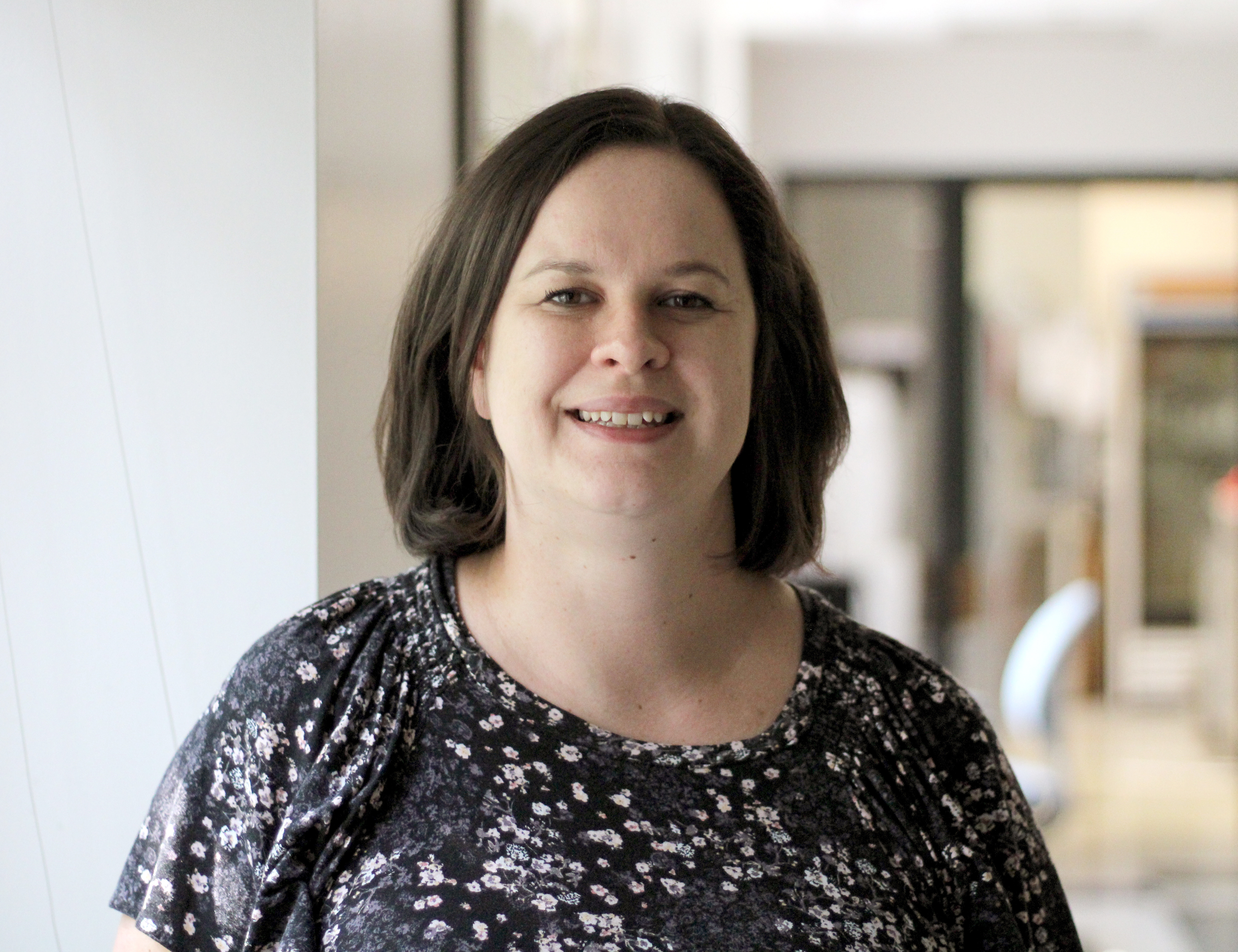
Christie Herd, a postdoctoral fellow in the Alexander Franz Veterinary Pathobiology lab won the Best of Show Award for her image of the La Crosse Virus in mosquito ovaries.
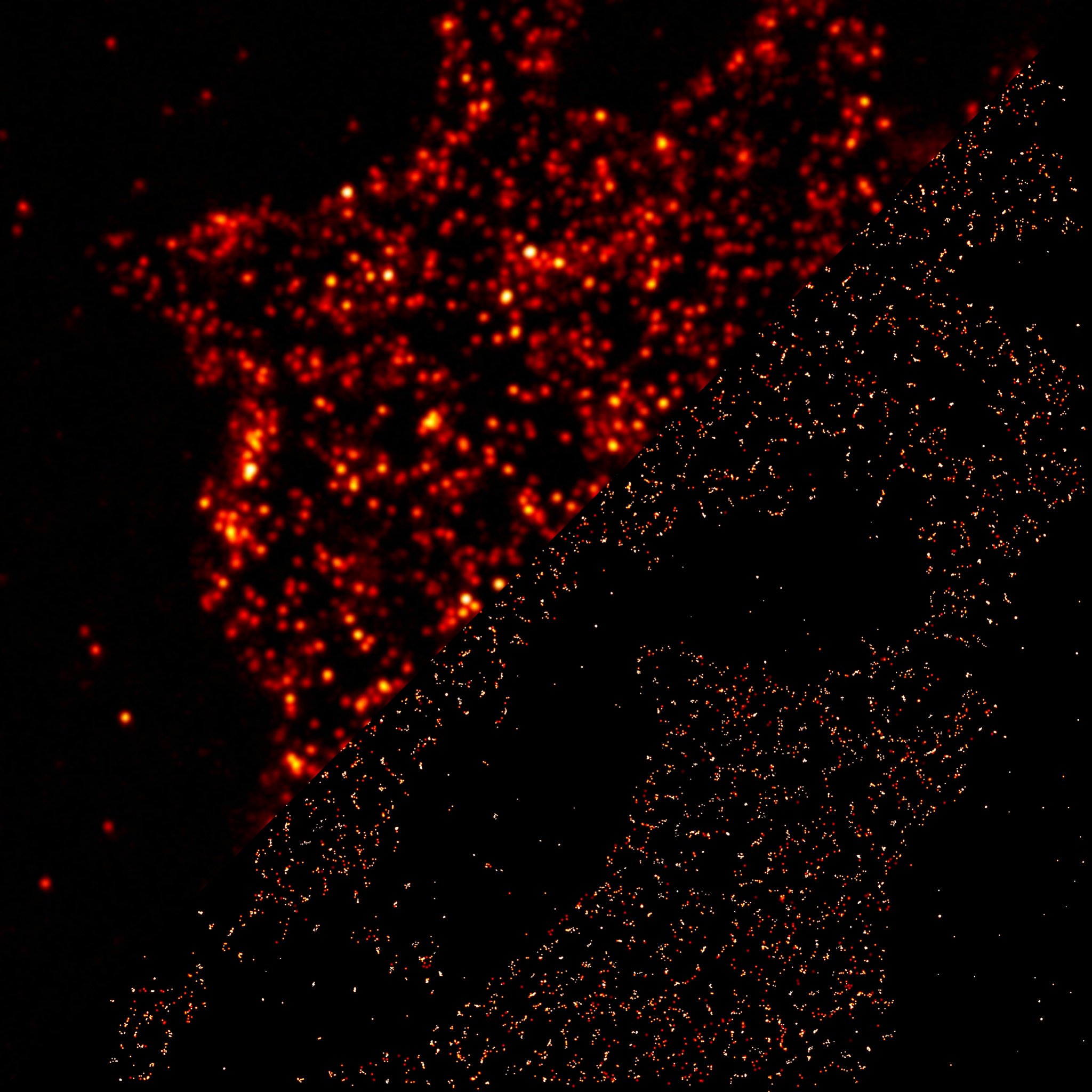
David Porciani, assistant research professor under the supervision of Bond LSC’s Donald Burke, won Director’s Award for Best Technically Challenging Image with his image showing epidermal growth factor receptors in two types of resolution.
Janlo Robil, a graduate student in the Bond LSC’s Paula McSteen lab won Experts’ Choice Award with his image of hormones in a corn leaf.
A wonderful surprise
Herd was “blown away” when she got her colorful image that won the Best of Show Award. Not only had she tried imaging other viruses with less success, she also worked on different La Crosse samples with no luck.
“That image in particular, I was not expecting to see because the day before I was there for three hours and had to quit,” Herd said. “So, when I saw that image, I was surprised because I did not think I would see that much detail.”
Herd’s image shows the La Crosse virus in developing eggs within mosquito ovaries. Herd uses La Crosse, a type of bunyavirus, as a model to study virus transmission from a female mosquito to her larvae to determine how viruses can remain transmitted within generations of mosquito populations. While La Crosse infects a small amount of humans a year, it transmits quickly, making it the perfect model to learn more about other bunyaviruses like Zika and Chikungunya.
“With bunyaviruses, they’re so multifaceted, and I like being able to research different aspects of them,” Herd said. “I just feel like they’re medically important.”
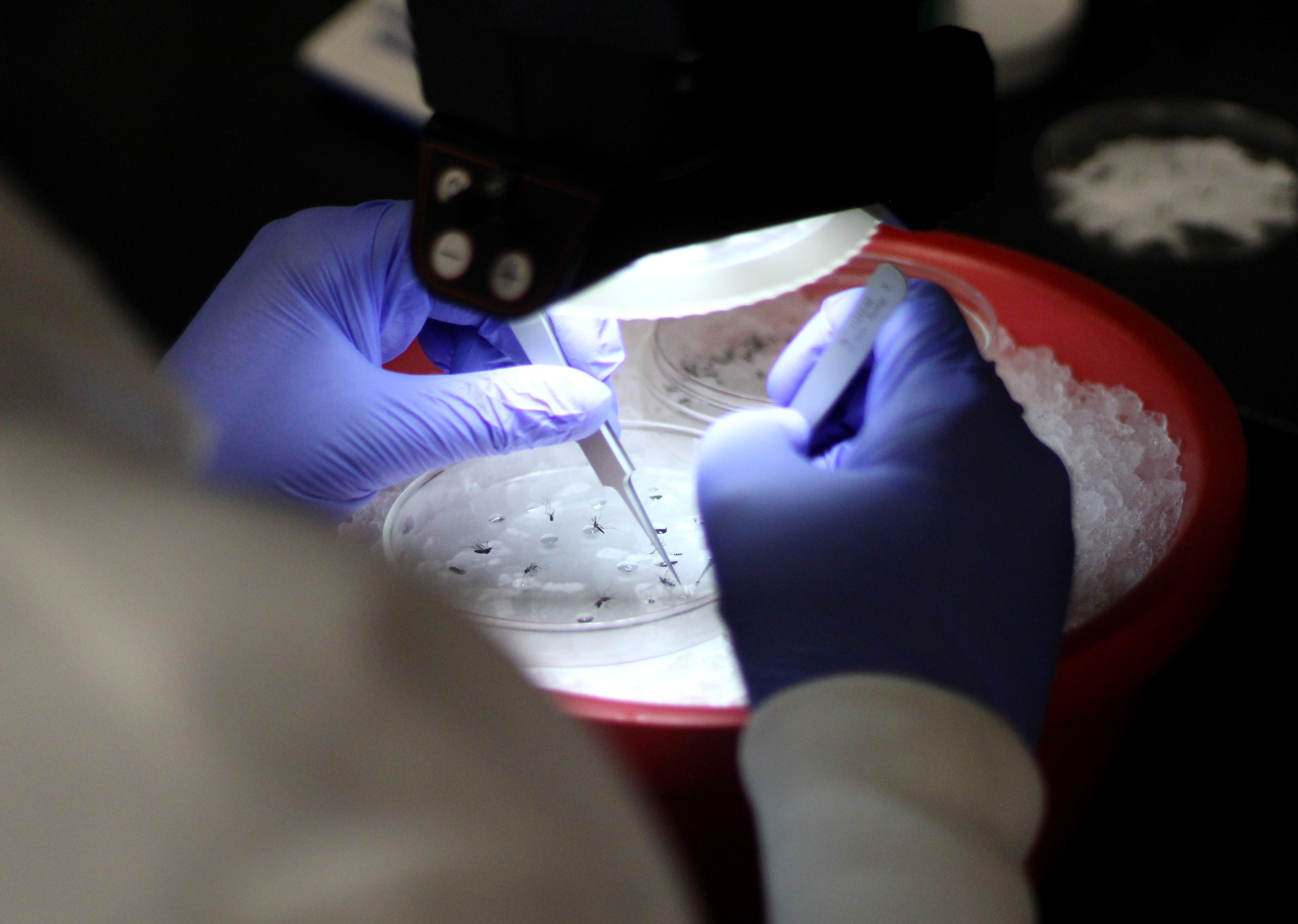
Herd’s image includes different colors to label different parts of the ovaries as well as the virus so she can see where in the ovaries the virus is traveling. Using a technique that allows her to see different focal planes, Herd can see where the virus is in every dimension, but it can be tricky to get high quality images.
“It does require a few hours of playing around, looking at the microscope,” Herd said with a chuckle. “It requires an investment of time, and sometimes there are days where it’s just not what you wanted and it doesn’t work … and you wasted days.”
Even with the challenges, the ability to see multiple dimensions of a sample at once is valuable for research projects like Herd’s.
“You just have to persevere and try again with a new set of samples,” Herd said.
From side project to passion
Although a deviation from his primary research, David Porciani’s project to image cell surface receptors slowly took over his focus.
“The biggest surprise was that I really had fun,” Porciani said. “This was not my primary project, but it became my primary project for a while.”
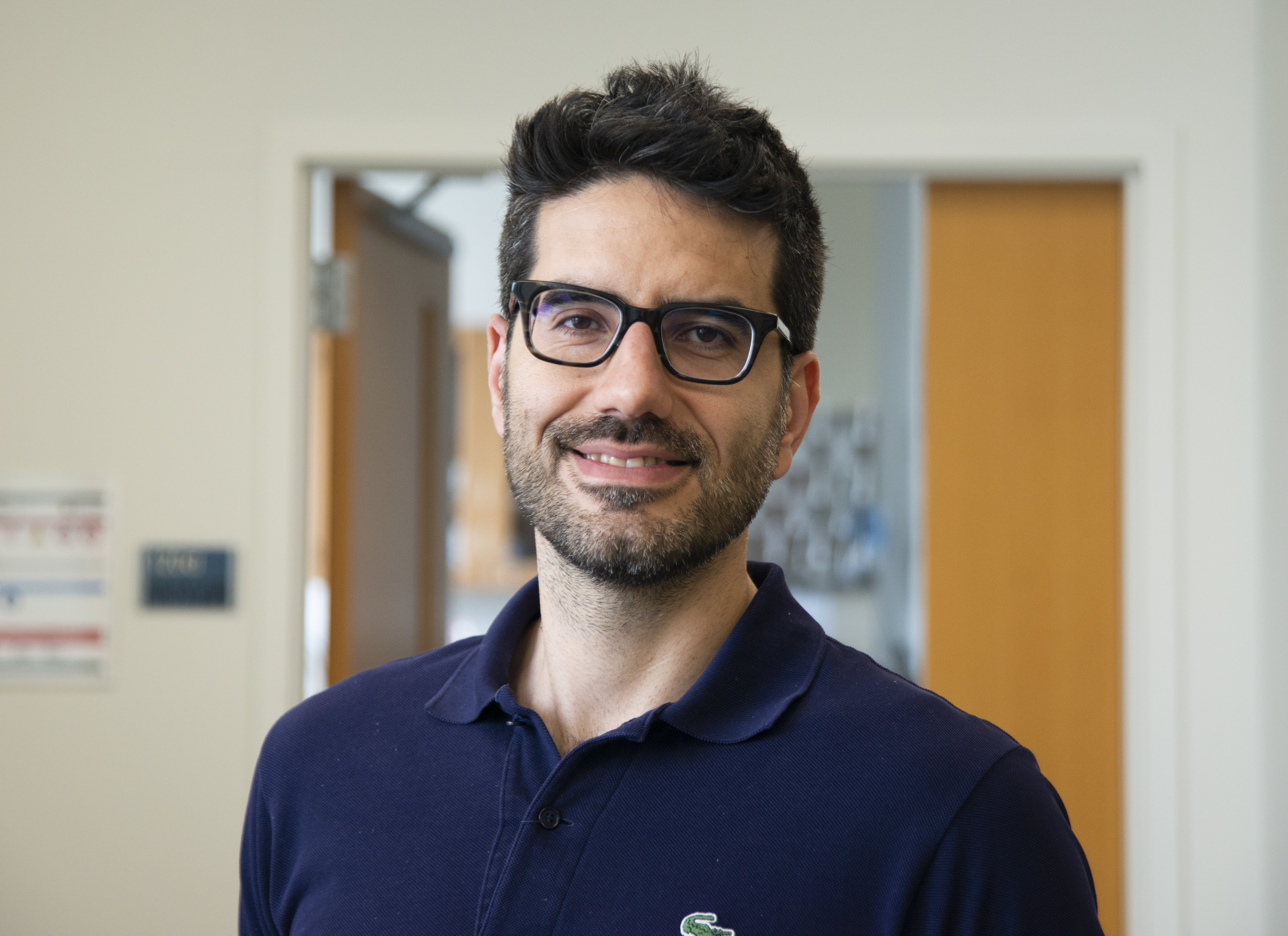
Porciani, an assistant research professor under the supervision of Donald Burke, studies molecules on the surface of cancer cells which are receptors for growth factors. These receptors act as a lock, with growth factors as a key. When the growth factors and receptors come together, the cells divide and create more cells. In lung cancer, there are more of these receptors, which can lead to uncontrolled tumor growth.
“This receptor, EGFR, has been widely studied,” Porciani said. “But for me there is definitely an interest because it’s one of the markers in lung cancer.”
Porciani tags those receptors with small molecules called fluorophores that glow under the light of the microscope, so he can see where the receptors are and how they move. However, the fluorophores cannot attach to the receptors alone, so he used aptamers — synthetic keys created by researchers that can bind receptor locks with specificity similar to the natural growth factors. Ultimately, the aptamers clip the fluorophores to the receptors.
“If you can follow the motion of the receptors, these receptors are kind of dancing,” Porciani said.
In his image, each dot is a different receptor made visible by the attached fluorophore.
However, fluorophores can become bleached, rendering them invisible after being exposed to the laser from the microscope for a while. If the synthetic keys, or aptamers, are still bound to those receptors, they cannot be imaged any longer. To address this, Porciani developed an aptamer which attaches to the receptor for a shorter time and then detaches so that even if the fluorophore bleaches, another aptamer can replace it and so receptors can be imaged for longer time.
“We engineered an aptamer with lower affinity that could work with this approach,” Porciani said. “By having lower affinity aptamers we can still determine localization of a high number of receptors and their motion.”
For his winning image, Porciani split the image to show the difference between single molecule resolution on the bottom right and a lower resolution image on the top left.
“With [lower] resolution, you don’t have a single molecule solution. If there are two molecules close together you will see them as just one single dot,” Porciani said. “But with the image, after software analysis with the image on the bottom right, then you have single molecule resolution.”
With this technique — made possible with microscopes at ALMC in Bond LSC — Porciani saw his efforts come together.
“At the beginning, I was just focused on the aptamer engineering from a high affinity aptamer to low affinity aptamer, and making them was the fun part to play with the structure,” Porciani said. “But when we started doing the imaging experiments at the Bond Life Sciences Center I realized that it was not just fun, but it was actually meaningful and this approach could have lots of biomedical applications.”
The artist’s touch
Although passionate about biology and microscopy, Janlo Robil decided to submit his image based on aesthetics.
“I chose this one because I am also a graphic artist, and I appreciate the color and composition,” Robil said.

Robil’s image shows a developing corn leaf with different colors labeling different plant hormone response proteins involved in stimulating growth. His unique image of an entire leaf, which is just under a millimeter in length, required piecing together images of sections of the leaf.
“About 0.75 millimeter, that’s still big in the microscope,” Robil said. “This is kind of difficult to make because it means that you need to tile several images [together] and sometimes it takes up to an hour just to [get] an image.”
The experiment requires planning ahead since the plants containing the fluorescent proteins must be crossed with plants with genetic mutations to determine the roles of the hormones in the leaf development.
To tag the plant proteins with fluorescent proteins requires planning ahead since the plants must be grown with genetic mutations.
“The beautiful image is actually a result of the expression of fluorescent proteins that are tagging the hormone response protein and also the hormone transport protein,” Robil said.
Initially from the Philippines where the agricultural staple is rice, Robil came to Mizzou interested in genetic mechanisms to make rice plants more productive. One way to enhance the rice plants is to make the rice leaves more similar to corn leaves, so Robil found interest in the McSteen lab’s project understanding the role of hormones in corn leaves.
“This project is perfect for me because I am studying the leaf and also integrating genetics,” Robil said. “And I love microscopy so much. I had quite a good amount of training starting in 2021 on confocal microscopy, and that’s why I was able to image this.”
![Christie_Herd_LACV_Ovaries[3528]](/wp-content/uploads/2022/05/picture-perfect-science-contest-highlights-best-microscopic-images-of-the-year.jpg)
![JMR_Life_Sciences_Week_Image_01[3346]](/wp-content/uploads/2022/05/picture-perfect-science-contest-highlights-best-microscopic-images-of-the-year-2.jpg)