Published on

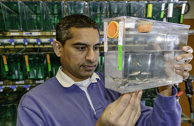
Bond LSC scientist Anand Chandrasekhar studies the zebrafish model to learn how motor neurons develop. These adult zebrafish lay eggs used to gain insight into how motor neurons arrange themselves as embryos grow into adults. Roger Meissen/ Bond LSC
Three thousand zebrafish swim circles in tanks located on the ground floor of the Bond Life Sciences Center, content to mindlessly while away their existence by eating their fill and laying eggs.
Despite their very basic higher functions, Bond LSC researcher Anand Chandrasekhar wants to understand how their brains work. More importantly, he wants to know how individual neurons end up ordered, all in the right place to support the animal’s automatic functions like breathing, swallowing and jaw movement.
This could one day lead to better understanding of specific neuronal disorders in humans.
“We are studying how cells end up where they are, and in the nervous system that’s especially critical because these neurons are assembling circuits just like in computers,” Chandrasekhar said. “If those circuits don’t form properly, and if different types of neurons don’t end up where they are supposed to, the behavior of the animal is going to be compromised.”
Zebrafish are a perfect model to study for many scientists.
Given plentiful food, adult zebrafish lay thousands of eggs that fall through a screen in the bottom of fish tanks to be collected. These eggs turn into new embryos that are nearly transparent, allowing for easy observation of the 3-4 mm fish under a microscope.

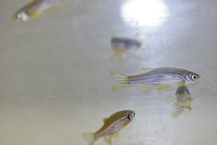
Zebrafish embryos are nearly transparent when born, making internal processes easy to observe. Only as they mature into adults like these do their scales develop pigment. Roger Meissen/ Bond LSC
Scientists have not only sequenced the zebrafish genome, but they’ve also inserted fluorescent jellyfish protein genes into the genome. This allows easy tracking of neurons.
“It’s called a reporter, and it’s used all the time to visualize a favorite group of cells. In our case, all of these green clusters of lighted up cells are motor neurons,” Chandrasekhar said. “Some groupings are shaped like sausages, some are more round, but each cluster of 50-150 cells sends out signals to different groups of muscles.”
These motor neurons are concentrated in the hindbrain. Akin to the brain stem, it controls the gills, breathing and jaw movement in these tiny fish. Genes controlling these functions are similar in zebrafish, higher vertebrates and humans despite their evolutionary paths diverging millions of years ago.
“The brain stem is the so-called reptilian brain that has changed very little, looking very similar in a lamprey or an eel all the way up to humans. All the elaborate brain structures you see in the cerebellum and the cortex are built up on top of that,” Chandrasekhar said. “Motor neurons from the brain stem send connections to muscles of the head including the gills, jaw and, in humans, the tongue, neck, voice box and muscles for breathing. We study those motor neurons and they undergo migration in many of these vertebrates systems.”

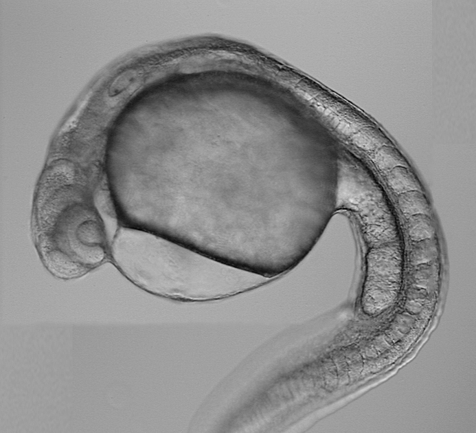
Zebrafish embryos measure only 3-4 mm seven days after they hatch. Chandrasekhar observes their motor neurons in this stage to discern how they migrate as the embryo develops. Courtesy of Anand Chandrasekhar.
This migration and signals that control it are what Chandrasekhar’s research is all about.
For example, his recent studies suggest that a protein called Vangl2 plays an important role in regulating movement of neurons through the zebrafish embryo’s matrix of tissue. Proteins like this are present in many organisms, from flies to fish to mammals.
“When I say a neuron is migrating in its environment, it’s actually pushing its way in between all these other cells,” Chandrasekhar said. “Cells in the environment of this migrating neuron secrete proteins that may diffuse away from the cell and bind to a receptor on a migrating neuron and then kind of beckon this neuron to keep moving.”
Chandrasekhar’s work contributes to a better understanding of how basic neuronal networks are created in development. That sort of knowledge could one day help with understanding the mechanistic bases of diseases like spina bifida, a nervous system disorder that results in muscle control problems due to the spinal cord not completely closing. Versions of this defect affect 1 in every 2,000 births, according to the National Institutes of Health.
“The significance of the work that we are doing is quite high for development,” Chandrasekhar said. “It is clear that even for the process of closing of the neural tube in the spinal cord and the brain, those cells closing the neural tube actually know left side from right side. The same kinds of mechanisms are going to be important and required whether you are talking about zipping together the neural tube or about allowing cells to squeeze between other cells to migrate and end up in a target position.”
Chandrasekhar’s work on Vangl2 recently published in the Feb. 2014 edition of Mechanisms of Development and the Oct. 2013 edition of Developmental Biology. Chandrasekhar is a professor of biological sciences in the Division of Biological Sciences and Bond LSC researcher.
Read more about his work at www.chandrasekharlab.net.